


A 32-year-old male presented to the emergency department for confusion, dyspnea, and a “white out” of his central vision over the preceding 24 hours. The patient had recently consumed a bottle of alcohol purchased overseas. Bloodwork revealed a severe metabolic acidosis (pH 6.90) and a critically high methanol level of 28.9 mmol/l. Shortly after presentation, the patient went into respiratory failure and became comatose. He was intubated and admitted to the ICU.
CT head obtained shortly after presentation was normal (not shown). MRI head was most significant for symmetric foci of diffusion restriction limited to the dorsolateral putamina (Figure 1A). These foci showed high signal on fluid attenuated inversion recovery (FLAIR) (Figure 1B). There was no associated susceptibility change on gradient echo to suggest hemorrhage (Figure 1C). There were no abnormalities elsewhere.
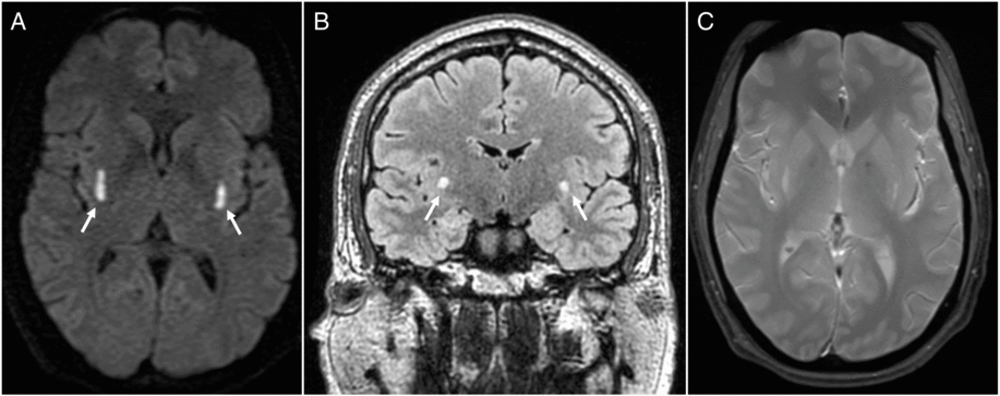
Figure 1: Focal and symmetrical signal change of dorsolateral putamina (arrows) is readily apparent on DWI (A) as well as FLAIR (B). There is no susceptibility artifact to suggest associated hemorrhage on multiplanar gradient-recalled echo (C).
The patient received two rounds of hemodialysis and was treated with fomepizole. He was extubated after 5 days and discharged 1 day later with a normal neurologic examination. Visual symptoms resolved over the following month and there was no evidence of optic neuropathy at follow-up with ophthalmology. The patient was lost to further follow-up and there was no repeat neuroimaging.
Previously reported MRI findings in methanol toxicity include high T2 signal suggestive of necrosis of the lentiform nucleus with predilection for the putamen. Reference Pelletier, Habib, Khalil, Salamon, Bartoli and Jean1 There may be necrosis of lobar white matter, classically with sparing of subcortical fibers, as well as hemorrhagic transformation. Reference Izumi, Ano, Komura, Hayashi and Adc2 Diffusion weighted imaging (DWI) findings have been less extensively described but include diffusion restriction of necrotic tissue. Reference Vara-Castrodeza, Peréz-Castrilĺn and Duẽas-Laita3 CT findings mirror MRI changes; early on, CT may be normal. Reference Taheri, Moghaddam, Moharamzad, Dadgari and Nahvi4
Our case is unique as abnormalities are limited to very focal signal change of the putamina with no white matter involvement and no hemorrhage. This in stark contrast to the majority of cases in the neuroimaging literature which describe extensive lentiform and lobar necrosis frequently with poor clinical outcomes. Reference Pelletier, Habib, Khalil, Salamon, Bartoli and Jean1 – Reference Vara-Castrodeza, Peréz-Castrilĺn and Duẽas-Laita3 Rather, in this case, other structures that have been implicated in methanol toxicity were normal, including the globus pallidus, thalamus, and temporal lobes. We report a distinct and comparatively subtle pattern of signal change featuring prominent diffusion restriction focally of the dorsolateral putamina. There are few cases in the literature of similar pattern of T2 change, Reference Hantson, Duprez and Mahieu5 – Reference Chattopadhyay and Chandra7 although these case reports do not present diffusion findings.
Decreased level of consciousness, respiratory failure, Reference Hovda, Hunderi, Tafjord, Dunlop, Rudberg and Jacobsen8 severe acidosis, and delay between ingestion and presentation Reference Hassanian-Moghaddam, Pajoumand, Dadgar and Shadnia9 have been associated with poor clinical outcomes in methanol toxicity. Despite satisfying all these criteria, this patient’s clinical course was quite favorable. We propose that, alongside clinical and laboratory status, early neuroimaging may have prognostic value in methanol toxicity.
The differential for bilateral lesions of the basal ganglia includes toxic, metabolic, genetic, and neurodegenerative conditions. Reference Hegde, Mohan, Lath and Lim10 However, MRI abnormalities, including on DWI, of the dorsolateral putamina may be highly suggestive of methanol toxicity, even if limited in extent. Early MRI should be considered as a supplement for prognostication in these patients.
Disclosures
The authors have nothing to disclose.
Statement of Authorship
All authors contributed to drafting and editing the manuscript.

